Section 8: Conventional Tomography
Introduction
This chapter provides information on the requirements and procedures for quality assurance testing of conventional tomographic x-ray machines. Although tests specifically pertaining to tomographic units are not mentioned in the Healing Arts Radiation Protection Act, the HARP Regulation 543, Section 8(10) require that quality assurance tests be performed every 6 months or upon servicing of every medical x-ray machine.
Note:
Several phantoms are available, therefore whichever is used follow the manufacturer's direction.
Mandatory HARP Tests
Mechanical Stability
Section Level (Fulcrum Accuracy)
Section Thickness
Suggested Tests
Level Incrementation
Exposure Angle Resolution
Patient Entrance Exposure
|
Note: All tests performed on conventional radiography systems must also be performed
Tomography involves controlled movement of the x-ray tube and film or cassette holder relative to one another during an exposure to produce a thin-section image. With linear tomographic movement, the tube and film move in opposite directions. The tube and cassette are joined by a rod or bar so that they move at the same speed and they maintain their relative positions. The pivot point on this metal bar is known as the fulcrum.
The anatomical part which is located at the same level as the fulcrum will not be blurred during the exposure, however all structures found above and below the fulcrum will be blurred.
It must be possible to examine structures at various levels within a patient. Some units allow the actual fulcrum to remain at a constant level and move the patient up and down (on a motordriven variable height table). Other units leave the patient at a constant level and allow the raising and lowering of the tube and film holder assembly.
The thickness of the tomographic section is directly related to the exposure angle (the angle produced by the tube movement). A small exposure angle produces a thick slice or section. A large exposure angle produces a very thin section.
Add-on tomographic systems generally require considerably more attention and service at shorter intervals than the sophisticated complex motion tomographic units, assuming that the same number of cases per day are done on each unit.
Performance criteria and tolerance limits for tomographic quality control tests vary somewhat with the type of tomographic unit. The unit's performance at the time of acceptance testing will set the baseline standard. For a typical tomographic unit, the following standards are provided as a guide to evaluate acceptable performance.
1.
Fulcrum Height Accuracy
a. Section level:
The agreement expected between the indicated and measured section levels varies somewhat, depending upon the type of tomographic unit. In all cases, however, agreement to within +/- 5 mm should be achieved. In measurements of this characteristic the level setting should always be approached from the same direction.
b. Level Incrementation:
In incrementing from one tomographic section to the next, level position should be reproducible to within +/-2 mm. In measurements of this characteristic, the level setting should always be approached from the same direction.
2.
Thickness of Cut
a. Section Thickness:
This characteristic varies with the type of tomographic motion and the exposure angle and uniformity. It is recommended that tolerance limits be established for each particular unit from images compared from one set of quality control measurements to the next. In measurements of this characteristic, the level setting should always be approached from the same direction.
b. Exposure Angle:
Indicated and measured exposure angles should agree to within +/-5 degrees. For units employing symmetric motion at wide angles, the symmetry of exposure angle should be within +/-5 degrees with respect to the midline.
3.
Mechanical Stability
The density of the image pattern on the resultant film from the pinhole test should be nearly uniform and straight. The image should reveal no unexpected overlaps, inconsistencies of exposure, or asymmetries in motion.
4.
Spatial Resolution
Most tomographic units should depict a 40 mesh screen pattern, ie. 40 holes per inch.
5.
Patient Entrance Exposure
In making exposure measurements, care should be taken to ensure that the dosimeter is positioned in the x-ray beam during the entire exposure. Quantitative criteria are unavailable for the values of PEE expected for tomography. Facilities should set their own baseline standards.
Mechanical Stability and Tube Angle (Pinhole Test)
Equipment
- One rectangular sponge approximately 5 cm in height.
- One lead aperture plate, 4 x 4 x 1/8 inch with a 1/16 inch hole in the centre.
- One 18 cm by 24 cm loaded cassette.
Procedure
1. Position the cassette in the bucky.
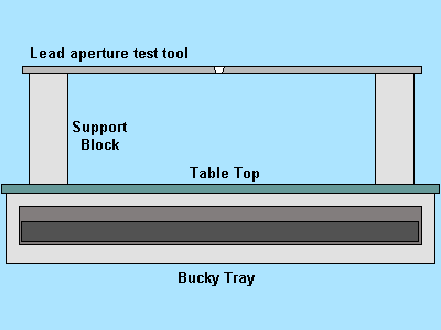
2. Place the lead aperture plate on top of the sponge and position on tabletop. Using the x-ray tube centering device, centre the x-ray tube over the plate. The hole in the lead aperture plate must coincide with the central ray of the x-ray field. (See Figure 8 - 1.)
3. Collimate the x-ray beam to a 10 cm by 10 cm field size.
4. Select radiographic mode and expose the cassette using approximately 50 kVp and 5 mAs. Do not remove the cassette from the tray.
5. Select the most commonly used tomographic mode and cut thickness.
6. Select a cut level of 12 cm.
7. Expose the cassette in tomographic mode for the second time using approximately 50 kVp and 10 to 20 mAs. (The technique will depend on type of film/screen & grid used.)
8. Process the film.
FIGURE 8-1 LEAD APERTURE TEST TOOL SETUP
 Evaluate as follows:
Mechanical Stability
The tomographic image of the hole in the aperture plate is a radiographic reproduction of the trajectory of the x-ray tube during the tomographic exposure. The black dot within the radiographic image indicates the centre of the trajectory (step 4).
For the linear tomographic mode, the black dot must appear within the centre third of the trajectory. If the black dot is outside the centre third of the trajectory, the angulation of the x-ray tube (tomographic arc) is not uniform and the system should be serviced.
The radiographic image should be evaluated to determine stability of the tube motion. The pattern reproduced on a linear system should be uniform in density and described as a straight line, not a wobbly one.
For pluridirectional systems, the images should be evaluated for completeness of the tomographic motion and degree of exposure overlap. Closure of all motions should be complete exhibiting no open gaps. Overlap should not exceed 20 degrees.
Should the test images reveal any large non-uniformities in the density of the projected trajectories (a waviness of the trace, incomplete closures or excessive overlaps) the cause should be determined and the system serviced.
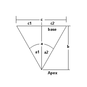
Calculation of Exposure Half-Angle Used in Linear Tomography
Measure, with a centimetre ruler, the length of the tracing on the radiograph. Construct a triangle with this measurement as the base. See Figure 8-2. Calculate (C/2b) mathematically. The exposure half-angle can be calculated as arctan (C/2b). Also refer to HARP Guidelines Page 145, Table 5.5.
b = Distance from apex to base of reconstructed triangle.
C = Measured with centimetre ruler on test film.
FIGURE 8-2 Calculation of Exposure Angle
 Fulcrum Height Accuracy
Equipment
- One 45 degree wedge sponge
- One collimation template or alternative tool
- One loaded 18 cm by 24 cm cassette
Alternative Equipment
If a collimation template is not available, a simple tomographic test phantom can easily be manufactured within a facility by following the steps listed below:
1. Cut a 30 degree - 60 degree - 90 degree triangular shaped piece from a 2 x 4 inch piece of wood.
2. Attach a copper window screen of #40 mesh to the angled surface of the wood block.
3. Attach a small piece of copper wire or a straightened paper clip to the screen halfway up the block.
Figure 8-3 Tomographic Test Tool
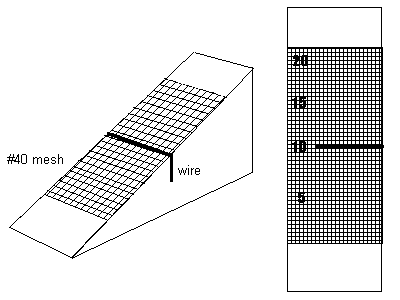
4. Attach lead numbers to indicate the cut levels.
5. Attach a thin lead sheet such as those found in old cassettes or cardboard film holders to the base of the wood block. This will some attenuation in order to get proper dentsity the on resultant film.
See page 138 of the Healing Arts Radiation Protection Guidelines for a more detailed explanation of the tomographic test tool construction.
Procedure
1. Place the cassette in the cassette tray so that the long dimension of the cassette is parallel to the long dimension of the x-ray table.
2. Tape the collimation template to the sponge at the 45 degrees angle. With the x-ray unit centering device, or with the tube perpendicular to the table, centre the template so that the central ray passes through the centre of the plate. For linear tomography, the wedge sponge must be
positioned perpendicular to the direction of motion. For non-linear (i.e circular, hypocycloidal, elliptical) the sponge can be placed parallel with the tube axis. Or position the alternative tool in place.
3. Adjust the collimation to provide a 5 cm by 5 cm field.
4. Measure the distance from the tabletop to the centre of the template or to a level on the
alternative tool and set this level on the cut level indicator.
5. Select the most commonly used tomographic mode and cut thickness.
6. Use technique factors of approximately 50 kVp and 5 to 10 mAs, and expose the film.(Depending on the film/screen speed.)
7. Develop film.
Evaluate as follows:
A properly functioning tomographic unit will produce one sharp area located at the indicated plane of cut if the fulcrum is accurate. This should be within 5 mm of the setting.
If using the alternative tool, in addition to above, the mesh area at the fulcrum height should be clear.
Erratic motion of the tomographic unit will produce several areas that appear to be sharp at different levels with blurred areas in between. Service is required.

